|
Getting your Trinity Audio player ready...
|
Neuroendocrine cancer of the gallbladder and bile ducts is very rare. It develops from neuroendocrine cells in the biliary system and can be a neuroendocrine tumour (NET), which is usually slower growing, or a neuroendocrine carcinoma (NEC), which tends to grow more quickly.
Neuroendocrine cancer in this part of the body can be difficult to diagnose because it may cause no symptoms at first, and symptoms that do occur can be similar to those caused by more common gallbladder or bile duct conditions.
This page explains more about neuroendocrine cancer of the gallbladder and bile ducts, including possible causes and symptoms, diagnosis, treatment and follow-up care. It also includes information about living with your diagnosis and where to find further information and support.
Use the links in the box to find the information that is most relevant to you.
What is neuroendocrine cancer of the gallbladder and bile ducts?
Neuroendocrine cancer is a type of cancer that begins in the neuroendocrine cells, which are found throughout the body. These cells have the ability to release hormones that can regulate many body functions. When they become cancerous, their growth rate and normal function is disrupted.
There are two main types:
- Neuroendocrine tumours (NETs) – usually slower growing
- Neuroendocrine carcinomas (NECs) – less common and faster growing
Neuroendocrine cancers arising in the gallbladder and bile ducts are extremely rare.
Treatment and outlook depend on factors including tumour type, grade, stage, differentiation and whether the cancer has spread to other parts of the body.
About the gallbladder and bile ducts
The biliary system and gallbladder work together to produce, store and release bile, a fluid that helps the body digest fats.
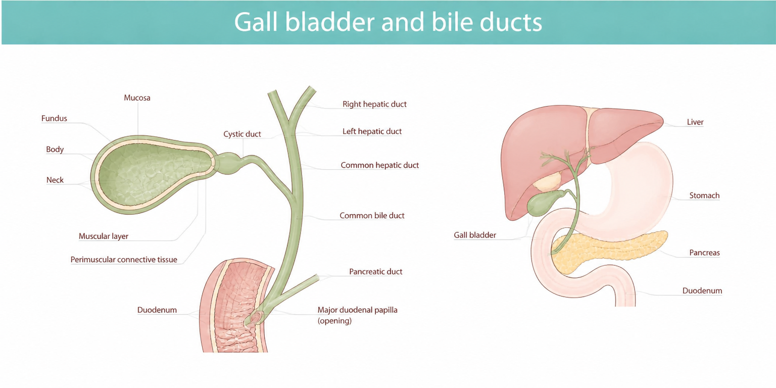
The biliary system (biliary tree)
The biliary tree is a network of tubes (ducts) that transport bile from the liver to the small intestine.
The main components include:
- Right and left hepatic ducts: drain bile produced by the liver.
- Common hepatic duct: formed when the right and left hepatic ducts join together.
- Cystic duct: connects the gallbladder to the biliary system.
- Common bile duct: formed by the union of the cystic and common hepatic ducts. It passes through the pancreas before emptying into the small intestine (duodenum).
- Sphincter of Oddi: a muscular valve that regulates bile flow into the intestine.
The gallbladder
The gallbladder is a small pear-shaped organ located beneath the liver. Its main functions are:
- Storing bile between meals
- Concentrating bile by removing water
- Releasing bile during digestion, particularly after eating fatty foods
When food enters the small intestine, hormones signal the gallbladder to contract and release bile through the bile ducts into the intestine.
What does bile do?
Bile contains water, bile salts, cholesterol and bilirubin.
The primary role of bile is to break fats into tiny droplets, allowing digestive enzymes to process and absorb them more effectively. Bile also helps remove waste products from the body.
Watch our video about the digestive system below.
How does neuroendocrine cancer of the gallbladder and bile ducts develop?
Neuroendocrine cancers develop when neuroendocrine cells acquire genetic changes that disrupt their normal growth and behaviour. Instead of growing and dying in a controlled manner, these cells continue to multiply and form a tumour.
Primary neuroendocrine cancers of the gallbladder and bile ducts are very uncommon, accounting for less than 1% of all neuroendocrine cancers.
The exact cause remains unclear. Possible contributing factors include:
- Chronic inflammation of the gallbladder
- Long-standing gallstones
- Changes in the gallbladder lining (metaplasia)
- Transformation of existing gallbladder cancers
- Abnormal development of stem cells within gallbladder tissue
Researchers continue to investigate the molecular changes involved in these cancers. Some studies suggest abnormalities involving:
- Epidermal growth factor receptor (EGFR) signalling pathways
- vascular endothelial growth factor (VEGF)
- Genes involved in tumour growth and blood vessel formation
Most cases occur sporadically and are not inherited.
However, if you have a family history of cancer (especially in those under 50), neuroendocrine cancer or a known hereditary cancer syndrome, it is important to discuss this with your specialist team.
Signs and symptoms
Many people have no symptoms during the early stages of disease. When symptoms occur, they are often non-specific and can resemble more common gallbladder conditions such as gallstones or inflammation.
1. Symptoms caused by the tumour itself
Symptoms may include:
- Pain or discomfort in the upper right abdomen
- Nausea
- Vomiting
- Abdominal bloating
- Loss of appetite
- Fatigue
- Unexplained weight loss
- Fever
- Jaundice (yellowing of the skin and eyes): this may occur if the tumour blocks the flow of bile through the bile ducts
- Dark urine
- Pale stools
- Itching (pruritus)
2. Symptoms caused by hormone changes
Most gallbladder and bile duct neuroendocrine cancers are non-functioning, meaning they do not produce excess hormones. Rarely, functioning tumours may produce hormones leading to symptoms such as carcinoid syndrome. Symptoms of this may include:
- Flushing
- Diarrhoea
- Wheezing
- Sweating
- Palpitations
- Abdominal discomfort
Carcinoid syndrome is rare in gallbladder neuroendocrine cancers. If it occurs, it is mostly associated with a gallbladder/biliary NET that has spread to the liver.
Carcinoid crisis (rare) may occur in those who have carcinoid syndrome
Carcinoid crisis can happen during anaesthesia or tumour manipulation (biopsy, surgery or interventional radiology – e.g. embolisation or ablation treatments) or be triggered by mental/physical stress.
Symptoms may be similar to carcinoid syndrome but are much more severe. These include severe flushing often accompanied by palpitations or a fast, irregular heart rate, low blood pressure, confusion and breathing difficulty, and can be life-threatening.
Specialist teams plan care to reduce this risk.
If you are at risk of carcinoid crisis, carry a Carcinoid Crisis Card so healthcare professionals know how to manage your care in an emergency. You can order one from us for free.
3. Rare symptoms (paraneoplastic syndromes)
Very rarely, cancer can trigger symptoms caused by the body’s immune response to cancer.
These are uncommon but can sometimes cause serious health problems.
Watch our video: Paraneoplastic Syndromes – Explained
4. Symptoms related to treatment
Treatments can cause side effects, but not everyone experiences them.
Your team will talk through possible side effects and how they can be managed.
Tests and scans
Because gallbladder and bile duct neuroendocrine cancers are rare and often mimic other conditions, diagnosis can be challenging.
Investigations are guided by symptoms, tumour behaviour and primary site. A comprehensive assessment usually includes:
- Clinical examination
- Blood and urine tests
- Cross-sectional imaging
- Functional imaging
- Endoscopy
- Histopathology
Your specialist team may recommend a combination of the following investigations.
Blood and urine tests
These may include:
- Full blood count (FBC)
- Liver function tests (LFTs)
- Kidney function tests
- Vitamin B12, iron studies, ferritin, folate
- Chromogranin A (CgA)
- Urinary or serum 5-HIAA – a breakdown product of serotonin
- CA19-9
- Carcinoembryonic antigen (CEA)
- NT-ProBNP may be taken if you have a history of heart disease or carcinoid syndrome (NET only)
- Vitamin, mineral and trace element check
Other blood tests may be taken, but this will be decided on a case-by-case basis, guided by your symptoms and clinical expertise.
Note that tumour markers can sometimes be normal even in the presence of disease – and some may give abnormal results for non-cancer reasons.
Scans
Structural imaging:
- Ultrasound scan
- CT scan
- MRI scan including magnetic resonance cholangiopancreatography (MRCP)
- CT chest, abdomen and pelvis
Functional imaging:
- Gallium-68 DOTATATE PET/CT
- FDG-PET/CT
- Other somatostatin receptor imaging e.g. octreotide SPECT scan
Additional imaging:
- Echocardiogram (where indicated)
- Bone imaging (if spread to bone is suspected)
Watch our video: Your Guide to Scans
Endoscopy
Endoscopic retrograde cholangiopancreatography (ERCP) is a specialised procedure that combines a thin, flexible camera tube called an endoscope with X-ray imaging to diagnose and treat problems affecting the bile ducts, liver, gallbladder and pancreas.
Percutaneous transhepatic cholangiogram (PTC) is a specialised imaging procedure used to examine the bile ducts and identify blockages or narrowing that may be causing symptoms such as jaundice, itching or infections. During the procedure, a radiologist inserts a thin needle through the skin and into the liver to access the bile ducts. A contrast dye is then injected, allowing detailed X-ray images of the biliary system to be taken.
Histopathology
This means looking at cells and tissues under a microscope to understand disease. It helps confirm whether cancer is present, where it started, what type of cells are involved, and how quickly the cells are growing.
Your doctor will explain which biopsy is best for you and why. You will also have time to ask questions and give consent before the procedure
A combination of these investigations will help confirm your diagnosis and guide treatment decision-making.
Cancer gene changes and targeted treatments
Some neuroendocrine cancers, particularly NECs, may contain specific genetic alterations that can be targeted with treatment. For example PD-L1, DLL3 and other molecular alternations.
Genomic testing may help identify targeted treatment options or eligibility for clinical trials.
Treatment options
The aim of treatment is to provide the best possible outcomes, symptom control and quality of life.
Treatment recommendations depend on:
- NET or NEC diagnosis
- Tumour grade
- Tumour size
- Location within the bowel
- Whether disease has spread
- Your overall health and preferences
1. Monitoring (surveillance)
This may be referred to as ‘watch and wait’ and may be used following treatment to check how well the treatment worked and to monitor stable disease. When active treatment is not currently needed, monitoring is required.
This typically involves a clinical review, blood tests and further scans or investigations.
2. Surgery
Surgery is the most important treatment for localised disease and offers the best chance of long-term survival. Surgery may be appropriate when:
- Disease can be safely removed
- Symptoms can be improved
The aim would be to completely remove the tumour and nearby lymph nodes.
However in NETs or where surgery is required to treat an obstruction, complete removal of all disease may not be possible. But debulking or removing an obstruction can still provide benefit.
Types of surgery, depending on the tumour location, may include:
- Cholecystectomy: removal of the gallbladder.
- Extended cholecystectomy: removal of the gallbladder with an inch or more of adjacent liver tissue, regional lymph nodes, and potentially the common bile.
- Liver wedge resection: there are two types: non-anatomical wedge resection (wedge) and anatomic segment 4b+5 resection (4b/5).
- Bile duct resection: removal of the part of the bile duct containing the tumour. Followed by reconnecting the remaining bile duct to the small intestine, creating a new route for bile to flow from the liver into the digestive system. This operation is known as a Roux-en-Y hepaticojejunostomy.
- Lymph node removal
The aim is complete tumour removal (R0 resection) whenever possible.
Endoscopic or radiology interventions
If surgery is not possible, there are other procedures that may help to relieve symptoms and improve quality of life. These treatments do not remove the cancer but can help manage problems caused by blocked bile ducts.
A biliary stent is a small tube placed inside a narrowed or blocked bile duct to keep it open. This allows bile to drain normally, which can help reduce jaundice (yellowing of the skin and eyes), itching, infection (cholangitis) and/or abdominal discomfort. Stents are usually inserted during a procedure such as an ERCP but if this approach is not possible stenting may be undertaken using the PTC procedure.
The main aim of stenting is to relieve symptoms, improve quality of life, and help maintain bile flow when the cancer cannot be completely removed by surgery.
Other interventions
In some select situations, certain procedures may be used to try to slow tumour growth within the bile ducts and help keep the ducts open for longer.
- Radiofrequency ablation: uses heat delivered through a specialised catheter to destroy some cancer cells within the bile duct. This may help reduce tumour size and delay the bile duct becoming blocked again.
- Photodynamic therapy: combines a light-sensitive medicine with a laser applied during an endoscopic procedure. The treatment targets and destroys cancer cells, which may help shrink the tumour and improve bile drainage.
In neuroendocrine cancer of the gallbladder and bile duct these procedures would only be available as part of a clinical trial as neither are currently approved for NHS use.
3. Non-surgical treatments
NET treatments
- Somatostatin analogues (SSAs): used to control hormone-related symptoms and/or tumour growth.
- Targeted medical therapy (e.g. the mTOR inhibitor everolimus): these are targeted treatments that block a protein that cancer cells use to grow and divide, helping to slow or stop tumour growth.
- Chemotherapy: rarely used as a first treatment for low grade (G1-2) NETs but may be considered in G2-3 or if other treatments are not suitable or have stopped working. It may be given as tablets or through a drip into a vein. For example CapTem (capecitabine + temozolomide) tablets or, rarely, Streptozocin/5FU via a drip.
- Radiotherapy: external beam radiation therapy (EBRT) may be used, following surgery or to treat symptomatic bone metastases.
- Radioligand therapy (RLT, also known as PRRT or lutetium‑177): mainly used for metastatic or progressing disease. It is a targeted internal radiation treatment. It delivers radiation directly to cancer cells, usually given in four sessions spaced 8–12 weeks apart.
- Symptom control and supportive care: includes any treatment or support that helps you feel better, even if it is not directly treating the cancer. This may include medicines, dietary advice and/or psychological support. Techniques such as mindfulness or relaxation may help with pain, fatigue, stress, or anxiety.
- Clinical trial therapy: clinical trials are used to find out if new treatments and techniques are safer and more effective than the ones that already exist, or for discovering if existing treatments can be used in different, more effective, ways or combinations.
NEC treatments
- Chemotherapy: considered to be a first line treatment for NECs in inoperable disease. It may also be considered if surgery is not possible or after surgery – if it was incomplete or doctors think there is a high risk of recurrence. The main treatment is usually platinum-based chemotherapy, sometimes combined with etoposide (or irinotecan in certain cases). If the cancer comes back or grows, further treatment options may include reusing the first treatment or trying other forms of chemotherapy.
- Radiotherapy: external beam radiation therapy (EBRT) may be used to treat symptomatic bone metastases.
- Immunotherapy: clinical trials are looking promising in treating NECs, but it has yet to become a standard of care. Research is ongoing. Immunotherapy may be considered in NEC if available and accessible. They include immune checkpoint inhibitors, which help your immune system recognise and attack cancer by removing the ‘brakes’ that normally hold it back, and DLL3 inhibitors, which target the protein DLL3, frequently over-expressed in high-grade neuroendocrine cancers.
- Symptom control and supportive care: includes any treatment or support that helps you feel better, even if it is not directly treating the cancer. This may include medicines, dietary advice and/or psychological support. Techniques such as mindfulness or relaxation may help with pain, fatigue, stress, or anxiety.
- Clinical trial therapy: clinical trials are used to find out if new treatments and techniques are safer and more effective than the ones that already exist, or for discovering if existing treatments can be used in different, more effective, ways or combinations.
Diagnosis and living with neuroendocrine cancer of the gallbladder and bile ducts
A diagnosis of neuroendocrine cancer can bring questions, uncertainty and a range of emotions. Information and support are available to help you understand your diagnosis and navigate what happens next.
Just diagnosed with neuroendocrine cancer? Access information and support to help you navigate the early days after diagnosis.
Follow-up care
Follow-up is an important part of your ongoing care. It helps your healthcare team check how you are doing, spot any changes early, and support your physical and emotional wellbeing.
While expert guidelines offer a general plan for follow-up, your care will always be tailored to you and your specific type of neuroendocrine cancer.
At each appointment, your team will ask how you are feeling, review your symptoms and medications, and arrange blood tests if needed.
You may also be offered a holistic needs assessment (HNA). This is an opportunity to talk with your specialist nurse or doctor about any physical, emotional, practical, or social needs, so the right support can be put in place.
Your case should always be reviewed within a specialist neuroendocrine multidisciplinary team (MDT) to ensure the most appropriate follow-up plan. Follow-up is primarily guided by multidisciplinary expert consensus and depends on whether the tumour is a well-differentiated NET or a poorly differentiated NEC.
If you have had treatment to remove the cancer
For well-differentiated NETs (grade 1 or grade 2)
- Usually require long-term follow-up
- Scans are often performed every 6 to 12 months
- Follow-up may continue for beyond 10 years as these tumours can occasionally return many years later
Well-differentiated NETs (grade 3) may require closer follow up, depending on Ki67%, symptoms and disease behaviour.
Poorly differentiated NECs (high grade) require closer monitoring. Scans are often performed every 3 to 6 months during the first few years after treatment. Follow-up is generally more intensive because these cancers can grow and spread more quickly.
If surgery is not possible or the cancer has spread
For NETs (grade 1 or grade 2), regular scans are usually performed every 3 to 6 months to monitor the cancer and assess how well non-surgical treatment is working.
For NECs and grade 3 NETs, follow-up is usually more frequent, often every 2 to 3 months, particularly during chemotherapy or other active treatment.
Special considerations for bile duct and gallbladder tumours
Because these tumours affect the biliary system, your team will also monitor for:
- Blockage of the bile ducts
- Jaundice (yellowing of the skin or eyes)
- Signs of infection
- Problems with stents or drainage tubes, if these have been inserted
How you can be part of your care
You are an important member of your healthcare team. You can help by:
- Reporting new symptoms or any changes in current symptoms or overall health
- Asking questions if at all uncertain or you want more information about your diagnosis and/or plan of care
- Attending appointments made – and checking up with the team if you haven’t received an appointment according to your agreed plan of care.
- Let your team know of your preferences and wishes regarding care – what is most important to you – be clear on concerns and influences on decision-making.
Support and further information
We have worked with the International Neuroendocrine Cancer Alliance (INCA) to provide information resources in more than 10 different languages: these are available here.
Other sources of support including benefits/financial, stoma care and general cancer advice include:
- Cancer Research UK: provides comprehensive guides on diagnosis and living with CUP, alongside a nurse-staffed helpline and online peer chat spaces.
- Maggie’s Centres: run drop-in centres across the UK providing free practical, emotional and social support from professional specialists.
- Macmillan Cancer Support: Offers a free support line (0808 808 00 00), for general cancer advice and financial guidance.
Stories from our community
Hear from people connected to neuroendocrine cancer as they share their experiences, perspectives and insights – from living with the condition to caring for, treating and supporting those affected.
Remember
Remember: every person’s experience with neuroendocrine cancer of the gallbladder and bile ducts is different – there may be shared similarities, but each journey is unique.
Your healthcare team will support you every step of the way, guiding your treatment, care, and follow-up. We’re here alongside them, offering additional support and information whenever you need it.
The information on this page is based on available clinical expert guidelines and publications. It is for informational purposes only and should not be considered professional medical advice.