|
Getting your Trinity Audio player ready...
|
Phaeochromocytoma is a rare neuroendocrine cancer that develops in the inner part (medulla) of the adrenal glands. The adrenal glands sit above the kidneys and make hormones that help control important functions in the body.
Phaeochromocytoma can affect people in different ways. Some tumours produce extra hormones, which can cause a range of symptoms, while others may cause few or no symptoms. Tests and scans can help your healthcare team understand your individual cancer and plan the most appropriate treatment and follow-up care.
This page explains more about phaeochromocytoma, including possible causes and inherited conditions, signs and symptoms, how it is diagnosed and monitored, and the treatments you may be offered. It also includes information about living with phaeochromocytoma, taking an active part in your care, and where you can find further information and support.
Use the links in the box to find the information that is most relevant to you.
What is a phaeochromocytoma?
There are two types of cancer that start in the adrenal gland:
- Adrenocortical carcinoma (ACC)
- Phaeochromocytoma (also known as adrenal-based paraganglioma)
Adrenocortical carcinoma (ACC) is a rare endocrine cancer that starts in the outer layer of the adrenal glands (called the cortex).
Phaeochromocytoma is a rare neuroendocrine cancer that starts in the inner part of the adrenal gland – called the medulla.
This page focuses on phaeochromocytoma.
Your adrenal glands
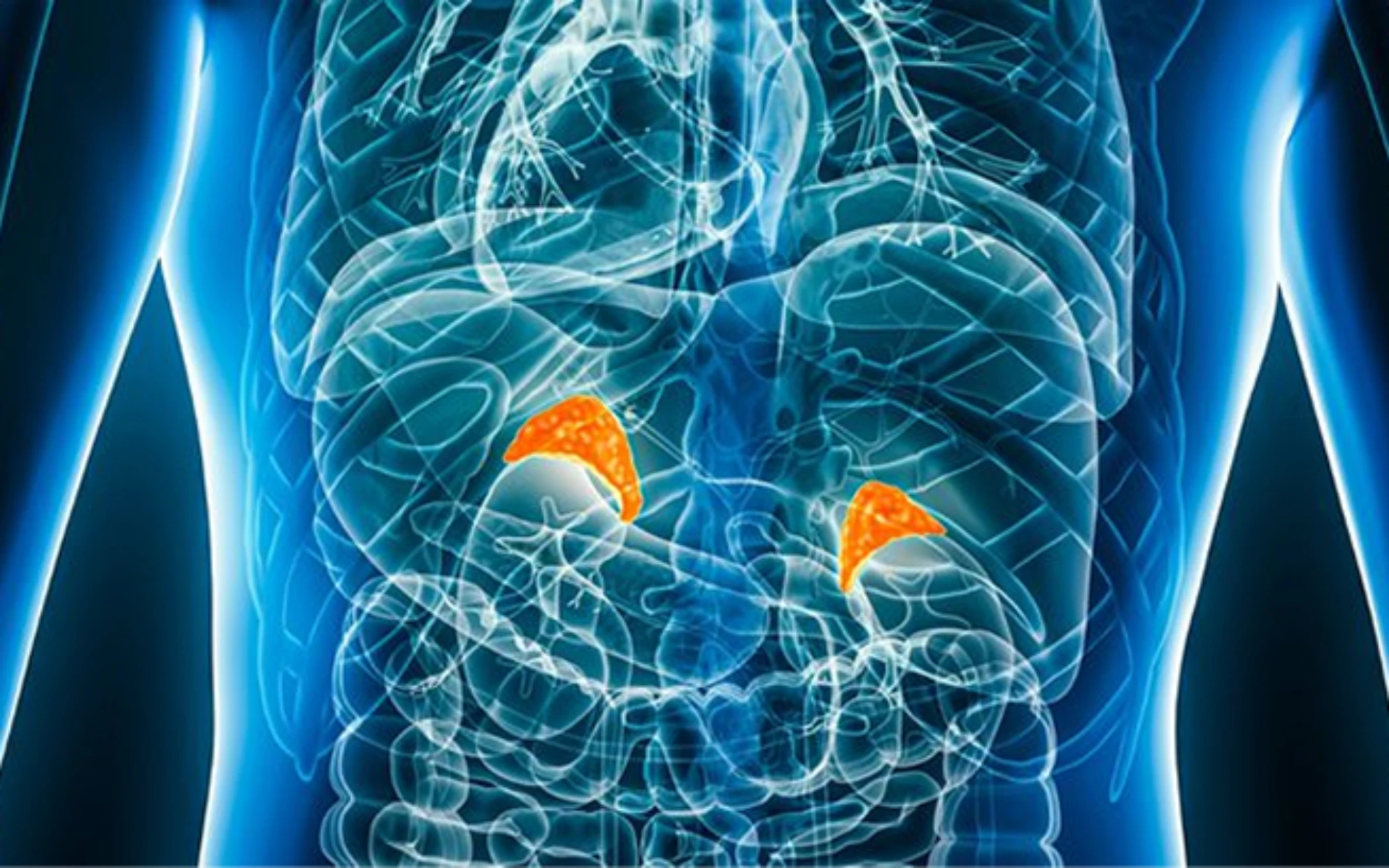
The adrenal glands are small organs that sit on top of your kidneys. They make hormones that help control important body functions such as:
- Blood pressure
- Energy levels
- Stress response
- Salt and water balance
When cancer develops in these cells, it can affect how hormones are made and released.
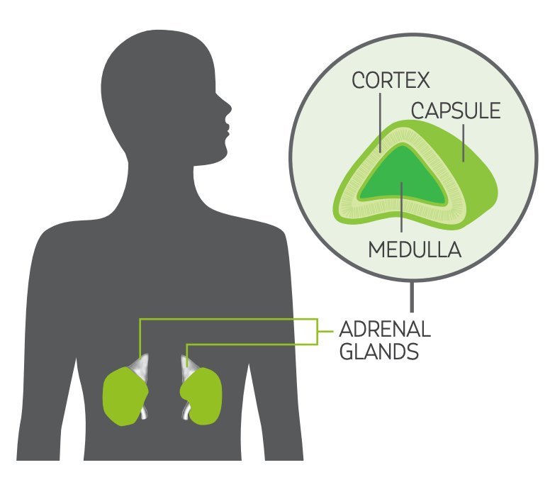
The adrenal gland
Each adrenal gland has three main structural parts:
- The capsule – a thin outer layer that surrounds and protects the adrenal gland
- The cortex (outer layer) – makes hormones such as cortisol, aldosterone, and small amounts of sex hormones
- The medulla (inner layer) – makes adrenaline (the ‘fight or flight’ hormone)
These hormones help your body respond to stress, control blood pressure, and manage how your body uses energy.
Phaeochromocytoma develops in the medulla of the adrenal gland.
How does phaeochromocytoma develop?
The exact cause of phaeochromocytoma is not yet fully understood. In some people and families, certain genetic changes (mutations) may increase the risk of developing this type of tumour.
Researchers are continuing to learn more about how the body works, how our environment may affect our health, and how genetic changes can lead to cancer.
This growing knowledge is helping to improve diagnosis and treatment and may lead to better ways to prevent cancer in the future.
Most phaeochromocytomas are sporadic, which means:
- They are not linked to an inherited condition
- They do not usually run in families
However up to 40% (4 in 10 people) with a phaeochromocytoma may have an inherited genetic condition. The risk may be higher (up to 50%; 5 in 10 people) when the tumour develops outside the adrenal gland (called a ‘paraganglioma’).
Inherited phaeochromocytoma conditions
Some genetic conditions are known to be linked with an increased risk of developing a phaeochromocytoma.
These include phaeochromocytoma and paraganglioma (PPGL) syndromes, multiple endocrine neoplasia 2 (MEN2), neurofibromatosis and von Hippel-Lindau (VHL).
Click on the drop-down options below to for patient support organisations associated with each inherited phaeochromocytoma condition.
PPGL syndromes
MEN-2
Neurofibromatosis
von Hippel-Lindau (VHL)
For those with either a phaeochromocytoma or paraganglioma, genetic testing may be offered to look for changes in a group of genes called succinate dehydrogenase (SDH) genes.
Experts recommend that everyone diagnosed with a phaeochromocytoma is offered genetic testing.
If a genetic change (such as an SDH mutation) is found, testing may also be offered to close family members, including parents, siblings (brothers and sisters) and/or children.
This can help identify others who may be at risk and ensure they receive the right monitoring and support.
Hereditary PPGL syndrome
This is caused by changes (mutations) in SDH genes (succinate dehydrogenase genes).
These genes normally help control how cells produce energy. When they do not work properly, it can increase the risk of certain tumours developing.
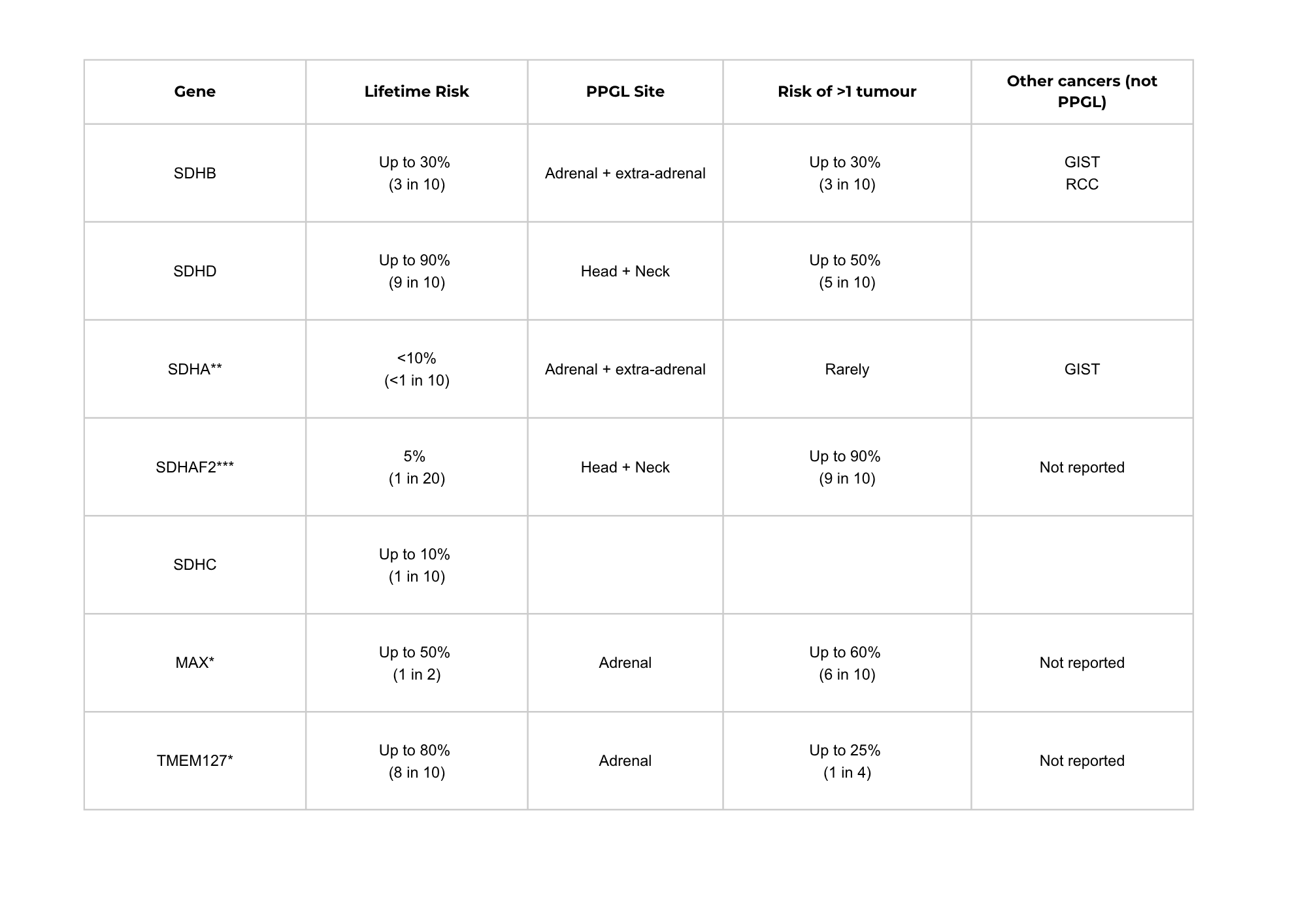
Table: PPGL risk and characteristics associated with different gene variants
Table notes:
‘PPGL site’ is the location in the body where phaeochromocytoma and paraganglioma associated with the gene may develop. ‘Adrenal’ means in the adrenal gland; ‘extra-adrenal’ means outside the adrenal gland.
PPGL: phaeochromocytoma and paraganglioma; GIST: gastrointestinal stromal tumour; RCC: renal cell carcinoma.
* Risk shown is for a mutation inherited from the father.
** Babies who inherit two SDHA mutations have a different genetic condition called Leigh syndrome. Further information on Leigh Syndrome is available at Metabolic Support UK
*** Risk shown is for a mutation inherited from the father. If inherited from the mother, children may be carriers and the mutation may be passed on to the next generation.
Risk of other cancers with hereditary PPGL syndrome
Not everyone with a family history, genetic condition or genetic mutation will develop a phaeochromocytoma, paraganglioma or other cancer.
People with a hereditary PPGL syndrome may have a slightly increased risk of developing other types of cancer, although this is still uncommon.
Gastrointestinal stromal tumours (GIST)
GISTs are rare tumours that develop in the wall of the stomach or bowel. They may grow slowly and not cause symptoms at first. They are very rare in people with hereditary paraganglioma-phaeochromocytoma syndromes. Routine screening is not usually needed, but they may sometimes be found during regular follow-up scans.
More information about GIST can be found at Gist Cancer UK.
Renal Cell Carcinoma (RCC)
Renal cell carcinoma (RCC) is a type of kidney cancer. It can also grow without symptoms in the early stages. If you are having regular scans as part of PPGL follow-up, any changes in the kidneys can often be picked up early.
More information about RCC can be found at Kidney Cancer UK.
If you have concerns about family history or genetic risk, your healthcare team can talk to you about whether genetic testing may be helpful.
Further information about PPGL syndrome and genetic conditions associated with phaeochromocytomas can be found by visiting our partner charities:
Signs and symptoms
Some people have no symptoms at first. Others notice changes before diagnosis.
Symptoms can happen for different reasons.
1. Symptoms caused by the tumour itself
Such as:
- Pain in the abdomen or side (flank)
- A feeling of fullness or a lump
2. Symptoms caused by hormone changes
Some phaeochromocytomas make extra hormones (called functioning tumours).
Symptoms can occur intermittently, often in episodes lasting only 15 minutes and may include some or all of the following:
- Headaches
- Dizziness
- Facial paleness (pallor)
- Excessive sweating
- Racing heart rate (palpitations)
- Panic attacks or a sense of doom
- Anxiety
- Weight loss
- Heat intolerance
- High (and rarely low) blood pressure (sustained or episodic)
- Nausea (with or without vomiting)
- Breathlessness
- Depression
- Lethargy
Many of the symptoms listed above are related to high blood pressure (hypertension) that can result from excess hormones released by this tumour.
Hypertension in patients with undiagnosed phaeochromocytomas can be very difficult to control; however, once a diagnosis is made, there are specific medications that can be prescribed to help with this.
Higher than normal levels of catecholamines (noradrenaline and adrenaline), can lead to catecholamine-induced hypertension (CIH) and more severely a CIH crisis, with a high risk of life-threatening complications (e.g. a stroke).
Once a phaeochromocytoma diagnosis is confirmed
If you have a phaeochromocytoma, you may need to avoid certain medicines that could make your symptoms worse. Your specialist healthcare team will advise you about which medicines are safe for you. Do not stop taking any prescribed medicines unless your healthcare team advises you to do so.
The Association for Multiple Endocrine Neoplasia Disorders (AMEND) has produced a patient information card that can be shown to a pharmacist or other healthcare professional to support medication prescribing.

AMEND patient information card
It is available from their website (www.amend.org.uk) or from your specialist team.
Seeking advice from a specialist team with expertise in managing phaeochromocytoma and paraganglioma is recommended to inform medications prescribing.
3. Rare symptoms (paraneoplastic syndromes)
Very rarely, phaeochromocytomas may trigger symptoms caused by the body’s immune response to cancer. These are uncommon but can sometimes cause serious health problems.
Watch our video: Paraneoplastic Syndromes – Explained
4. Symptoms related to treatment
Treatments can cause side effects, but not everyone experiences them.
Your team will talk through possible side effects and how they can be managed.
Adrenal insufficiency and adrenal crisis
If you have had surgery to remove one or both adrenal glands, you may be at risk of adrenal insufficiency and/or adrenal crisis.
- Adrenal insufficiency means the body cannot make enough steroid hormones, which are vital during illness or stress
- If not treated quickly, this can lead to an adrenal crisis, which is a medical emergency
- Risk increases during illness, surgery, injury or vomiting/diarrhoea: follow ‘sick day’ rules (available at The Addison’s Disease Self-Help Group)
- Always carry a Steroid Emergency Card if advised to
- Your specialist team will plan your care to reduce this risk
Tests and scans
Blood and urine tests
Expert guidelines recommend the following tests:
- Full blood count
- Liver and kidney function
Hormonal work-up:
- Glucocorticoid levels
- Dexamethasone suppression test
- 24-hour urine collection text for free urinary cortisol
- Basal cortisol (serum)
- Basal adrenocorticotropic hormone (ACTH) (plasma)
Sexual steroids and steroid precursors:
- Serum DHEA-S, 17=OH-progesterone, androstenedione and testosterone
- 17-beta-oestradiol (serum, men and postmenopausal women only)
- 24-hour urine collection to measure steroid metabolites
Mineralocorticoid levels:
- Potassium (serum), aldosterone/renin ratio (only in those with arterial hypertension with or without low potassium levels), catecholamine levels
- Normetanephrines, metanephrines and methoxytyramines (plasma)
- Alternatively; fractionated metanephrine production (24-hour urine collection)
Not all of these tests will be needed if it is already certain that you have phaeochromocytoma. Some are only required if the nature of the adrenal tumour is uncertain.
Scans
You may have:
- CT and/or MRI scans of the chest, abdomen and pelvis
- Metaiodobenzylguanidine (MIBG) or gallium dotatate PET scan
Watch our video: Your Guide to Scans
Biopsy (tissue sample)
Studying tissue under a microscope can be a helpful way to confirm a cancer diagnosis. It can give clues about where a cancer started, what kind of cancer cells exist, and how quickly or slowly they are dividing and growing.
Treatment options
Your care is individualised based on your tumour, health and preferences.
1. Monitoring (surveillance)
Often used after treatment or if treatment is not possible but intervention or supportive care may help with symptom management. Regular scans and check-ups help monitor for any changes.
2. Surgery
If possible, surgery aims to remove all of the tumour. There are two main types of surgery used in phaeochromocytoma:
Open adrenalectomy
This involves removing the adrenal gland through a cut (incision) in the abdomen. The size and position of the incision will depend on the size of the tumour and whether nearby structures also need to be removed. The cut may be horizontal, diagonal or vertical.
Laparoscopic adrenalectomy (keyhole surgery)
This is a less invasive operation using small cuts and a camera. However, it is not commonly used for phaeochromocytoma unless the tumour is very small. In most cases, an open operation is recommended to allow the surgeon to safely remove all of the cancer.
Your surgical team will discuss the most appropriate approach for you and explain what to expect before and after your operation.
Expert guidelines recommend that adrenal surgery should be carried out only by surgeons with appropriate expertise and experience.
Pre-operative considerations
Pregnancy and functional phaeochromocytoma
If you have a functional tumour and are planning a family, it is usually recommended to delay pregnancy until after treatment (such as surgery or radioligand therapy) and recovery.
If you become pregnant during, or just before, treatment, it is important to tell your doctor as soon as possible.
Your care will be planned by a specialist team, including your cancer team, obstetrician and midwife, to help keep both you and your baby safe.
Some tests and treatments are not suitable during pregnancy, as they may affect the unborn baby. These include:
- CT scans
- MIBG scans
- PET scans
- Radioligand therapy (RLT)
If surgery cannot be safely delayed until after the baby is born, and the tumour is suitable for keyhole (laparoscopic) surgery, this may be carried out during the second trimester (weeks 13–27) by an experienced specialist team.
In some cases, it may be safer to manage the tumour during pregnancy and plan surgery after the baby is born.
Further information and support is available from PhaeoPara Support UK.
Blood pressure control
High blood pressure (hypertension) caused by a phaeochromocytoma can be difficult to control, especially during surgery.
To reduce risks, you will usually be given medication called alpha-blockers (and sometimes beta-blockers) for at least 10 days before your operation.
These medicines help to lower blood pressure and reduce the risk of complications during surgery.
You may be prescribed these medications even if you have few or no symptoms, as they are an important part of making surgery safer.
Alpha-blockers before surgery
Common alpha-blockers include phenoxybenzamine or doxazosin. These are usually taken at home before surgery.
Some people may experience side effects, such as:
- Dry mouth
- Stuffy nose
- Feeling dizzy
- Feeling more tired than usual
Some men may notice changes during sex, such as difficulty ejaculating.
Your doctor or nurse will ask about any side effects to make sure the treatment remains safe and suitable for you.
In many cases, these medications can be stopped after the tumour has been removed.
Your healthcare team will guide you through each step and help you prepare safely for surgery. If you have any concerns, it’s always okay to ask questions.
3. Non-surgical treatment options
These may include:
- Somatostatin analogues: these injections are more commonly used in other neuroendocrine tumours to control hormone-related symptoms and slow tumour growth.
- Targeted therapy, e.g. sunitinib: sunitinib is a multikinase inhibitor (MKI), a type of targeted treatment that block several signals (called tyrosine kinases) that cancer cells use to grow and divide, helping to slow tumour growth.
These medicines can raise blood pressure, so in phaeochromocytoma it is very important that blood pressure is carefully controlled to make sure the treatment can be used safely.
- Chemotherapy: this is rarely used as a first treatment for phaeochromocytomas but may be considered if other treatments are not suitable or have stopped working. It may be given as tablets or through a drip into a vein. If considered, a combination of cyclophosphamide, vincristine and dacarbazine (CVD) is the most commonly used regimen, although temozolomide may also be considered.
- Radioligand therapy (RLT, also known as PRRT or lutetium‑177): this is a targeted internal radiation treatment. It delivers radiation directly to cancer cells, usually given in four sessions, spaced 8–12 weeks apart. It is mainly used for metastatic or progressing disease. At present, RLT with lutetium-177 does not have approval from the National Institute for Health and Care Excellence for phaeochromocytomas, but it may be available through clinical trials or other funding routes. You may be offered MIBG therapy instead, which is similar but uses a different radionuclide.
- Local therapies may include radiotherapy and/or interventional radiology, for example radiofrequency ablation, cryoablation, microwave ablation, (chemo-) embolisation. An individualised treatment approach is required.
- Radiotherapy: external beam radiation therapy (EBRT) may be used to treat symptomatic bone metastases.
- Symptom control and supportive care: this includes any treatment or support that helps you feel better, even if it is not directly treating the cancer. This may include medicines, dietary advice and/or psychological support. Techniques such as mindfulness or relaxation may help with pain, fatigue, stress or anxiety.
- Anti-hypertensives: these help to help control raised blood pressure. Your specialist team can advise and/or prescribe the type of medication best used in phaeochromocytomas. This may be different to the standard treatment used in non-hormonal-related hypertension.
- Clinical trial therapy: clinical trials are used to find out if new treatments and techniques are safer and more effective than the ones that already exist, or for discovering if existing treatments can be used in different, more effective, ways or combinations.
Immunotherapy treatments have yet to be proven effective in neuroendocrine tumours (NETs) and the precise role of immunotherapy remains unclear in the treatment of phaeochromocytomas. However, research is ongoing.
You can also find further information from our partner organisations:
Diagnosis and living with phaeochromocytoma
A diagnosis of neuroendocrine cancer can bring questions, uncertainty and a range of emotions. Information and support are available to help you understand your diagnosis and navigate what happens next.
Just diagnosed with neuroendocrine cancer? Access information and support to help you navigate the early days after diagnosis
Follow-up care
Follow-up is an important part of your ongoing care. It helps your healthcare team check how you are doing, spot any changes early, and support your physical and emotional wellbeing.
While expert guidelines offer a general plan for follow-up, your care will always be tailored to you and your specific type of neuroendocrine cancer.
At each appointment, your team will ask how you are feeling, review your symptoms and medications, and arrange blood tests if needed.
You may also be offered a holistic needs assessment (HNA). This is an opportunity to talk with your specialist nurse or doctor about any physical, emotional, practical or social needs, so the right support can be put in place.
Guideline follow-up recommendations for neuroendocrine cancers of the breast
After surgery you will usually have an early review:
- Around 2 to 6 weeks after surgery, with a clinical review and blood and/or urine tests
- This is repeated at around 12 to 16 weeks, often with scans such as CT or MRI (and sometimes functional imaging)
Ongoing follow-up usually includes:
- A review at 6 months
- Then yearly follow-up
For most people, follow-up continues for at least 10 years. However, if you also have a genetic condition, follow-up is usually lifelong.
Experts recommend the following tests and scans are done at or just before your follow up appointment:
- Blood and urine tests every year, including markers such as chromogranin A
- Scans every 1 to 2 years, unless your team feels they are needed more often
Because these tumours can sometimes spread to the bones, your doctors will choose scans that are sensitive to this risk.
For scans of the abdomen:
- An MRI scan is usually preferred
- A CT scan may be used if MRI is not suitable or available
If surgery was or is not possible or if the cancer has come back
Your follow-up plan will depend on your individual situation, including your general health.
Regular review is strongly recommended if you are receiving active treatment, have ongoing symptoms, or are assessed as being at higher risk of the cancer growing or returning
Hormone monitoring
For all people with phaeochromocytoma, regular checks are recommended to monitor hormone levels, alongside imaging.
Your follow-up plan will always be tailored to you. Your healthcare team will explain what to expect and when your tests and appointments will take place.
How you can be part of your care
You are a key part of your team. You can help by:
- Reporting new symptoms or any changes in current symptoms or overall health
- Asking questions if at all uncertain or you want more information about your diagnosis and/or plan of care
- Attending appointments made – and checking up with the team if you haven’t received an appointment according to your agreed plan of care
- Making your wishes and any concerns clear – let your team know what matters most to you.
Support and further information
We have worked with (the International Neuroendocrine Cancer Alliance (INCA) to provide information resources in more than 10 different languages: these are available here.
You can also find further information from our partner organisations:
Stories from our community
Hear from people connected to neuroendocrine cancer as they share their experiences, perspectives and insights – from living with the condition to caring for, treating and supporting those affected.
Remember
Remember: every person’s experience with phaeochromocytoma is different – there may be shared similarities, but each journey is unique.
Your healthcare team will support you every step of the way, guiding your treatment, care, and follow-up. We’re here alongside them, offering additional support and information whenever you need it.
The information on this page is based on available clinical expert guidelines and publications. It is for informational purposes only and should not be considered professional medical advice.