
|
Getting your Trinity Audio player ready...
|
Neuroendocrine cancers of the breast are rare cancers that develop in breast tissue. They include neuroendocrine tumours (NETs), which are usually slower growing, and neuroendocrine carcinomas (NECs), which tend to grow more quickly.
Neuroendocrine cancers of the breast can affect people in different ways. Breast examinations, scans and tests on a sample of the cancer can help your healthcare team understand your individual cancer and plan the most appropriate treatment and follow-up care.
This page explains more about neuroendocrine cancers of the breast, including possible causes and symptoms, diagnosis, monitoring and treatments. It also includes information about living with your diagnosis and where to find further information and support.
Use the links in the box to find the information that is most relevant to you.
What is neuroendocrine cancer?
Neuroendocrine cancer is a type of cancer that begins in the neuroendocrine cells, which are found throughout the body. These cells have the ability to release hormones that can regulate many body functions. When they become cancerous, their growth rate and normal function is disrupted.
There are two main types:
- Neuroendocrine tumours (NETs) – usually slower growing
- Neuroendocrine carcinomas (NECs) – less common and faster growing
This page looks at both NETs and NECs of the breast.
Your breasts
Everyone is born with breast tissue, no matter their gender.
Breasts change throughout life, from before birth, through puberty, adulthood and later life.
Before birth, breast tissue begins to form in the womb along lines from the armpits to the groin, and in rare cases extra nipples can develop.
During childhood, boys and girls have similar breast tissue with no major differences.
At puberty, hormones cause changes. In females, oestrogen and progesterone help breasts grow, forming milk ducts and milk-producing parts, with fat giving shape and size, and development can continue into the early twenties. In males, testosterone limits breast growth, so breasts remain small, do not produce milk, but can still be sensitive.
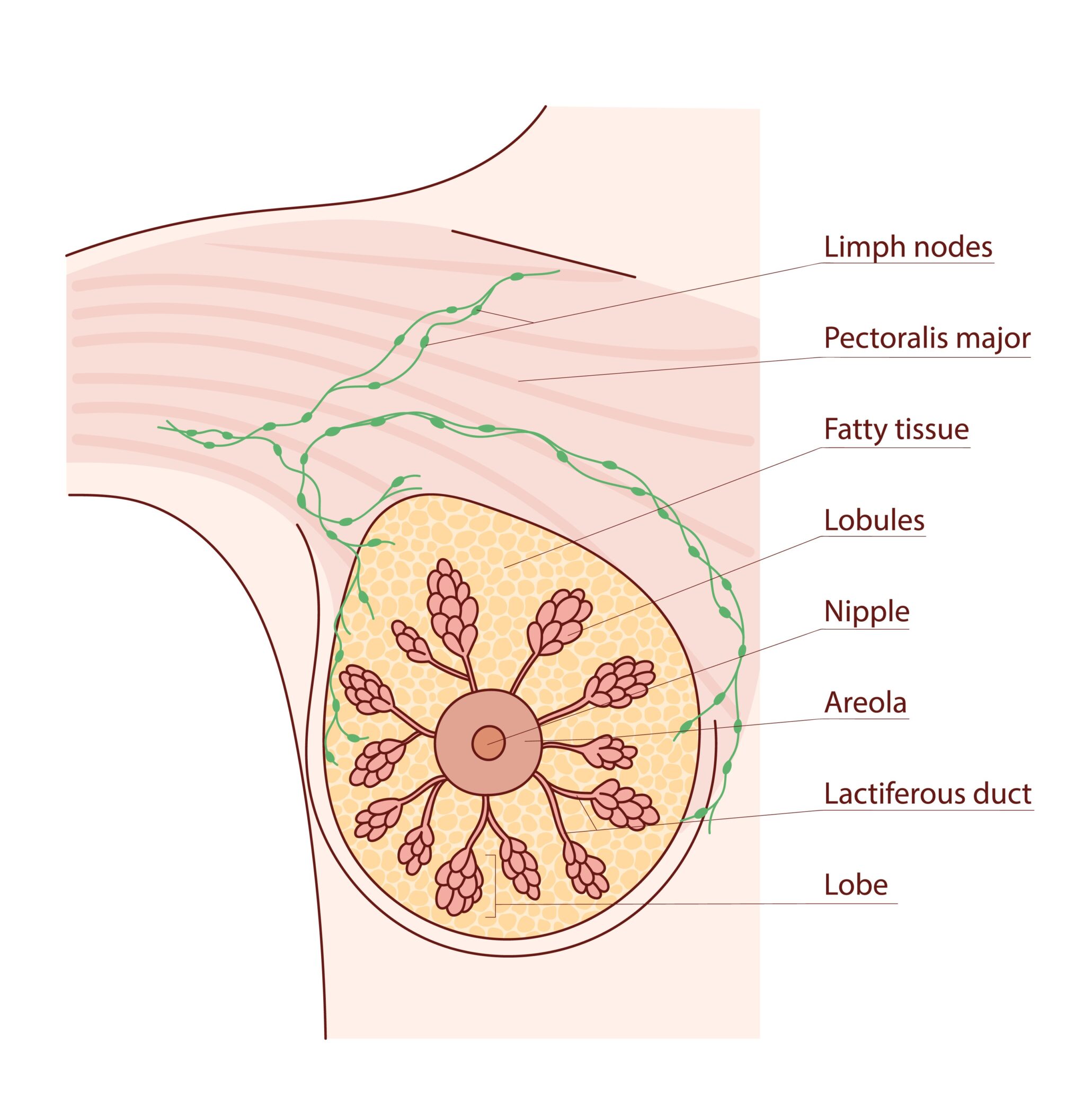
Structure of the adult breast
Breasts sit on the chest muscles. Tissue extends from the breast to the underarm area. Inside the breasts are:
- Lobes and lobules: make milk
- Ducts: carry milk to the nipple
- Fat and connective tissue: give shape and support
- Blood and lymph vessels: help with circulation and immune defence
Outer parts include:
- Nipple: where milk comes out; it can become firm with touch
- Areola: the darker are of skin around the nipple; it helps protects the nipple during breastfeeding
- Skin: similar to skin elsewhere on the body, with hair follicles and sweat glands
Breast size and shape varies naturally between people and can be affected by genes, hormones and body weight.
During pregnancy, breasts prepare to make milk. Milk-producing glands grow and become active, and nipples are lubricated to help breastfeeding.
During menopause, hormone levels fall, breast tissue becomes less dense, and milk-producing structures get smaller.
A note about gynaecomastia
Gynaecomastia is a condition where breast tissue becomes enlarged in men (male gender), sometimes called ‘man boobs’. It can happen at any age and may affect one or both breasts.
The main signs are breasts that look or feel larger than usual, and they may feel tender or sore. Common causes include normal hormone changes (such as during puberty or later life), being overweight, some medicines (like antidepressants or treatments for cancer), certain health conditions affecting the thyroid, liver or kidneys, and the use of drugs such as anabolic steroids.
Further information on gynaecomastia can be found on the NHS website.
How do neuroendocrine cancers of the breast develop?
For most people, there is no clear cause of breast NETs and NECs.
Most neuroendocrine tumours of the breast are not inherited, but a small number may be linked to genes passed down in families.
The main inherited risk comes from conditions that already increase the risk of breast cancer, such as changes in the BRCA1, BRCA2, TP53, or CHEK2 genes.
Rare inherited syndromes may also play a role. These include:
- Multiple endocrine neoplasia (MEN) syndromes, which increase the chance of developing some types of neuroendocrine or hormone-related tumours.
- Cowden syndrome, caused by a change in the PTEN gene, which raises the risk of breast and other cancers.
However, these conditions are very uncommon, and it is even more rare to find breast neuroendocrine cancers linked to them.
If you have a strong family history of breast, ovarian or neuroendocrine cancers, your healthcare team may suggest genetic counselling or testing through the NHS. This can help you understand your own risk and whether other family members might be affected.
Note that many genetic changes found in neuroendocrine cancers of the breast happen only within the cancer itself and are not inherited, so they do not affect your family’s risk.
Researchers are continuing to learn more about how the body works, how our environment may affect our health, and how genetic changes can lead to cancer.
This growing knowledge is helping to improve diagnosis and treatment and may lead to better ways to prevent cancer in the future.
How do hormone levels affect breast cancer risk?
The answer is not yet fully understood or known.
Klinefelter syndrome is a genetic condition affecting about 1 in 1,000 men, where an extra X chromosome is present. It can lower testosterone and increase oestrogen levels. Because of this, men with Klinefelter syndrome have a higher risk of breast cancer than other men, although the overall risk remains low. However It is not yet clear whether this risk extends to neuroendocrine cancers of the breast specifically.
Further information on Klinefelter’s syndrome and cancer can be found at the eXtra Klinefelter’s Syndrome Association.
For trans and non-binary people, hormone treatment and surgery can also affect risk.
Trans women (assigned male at birth) who take oestrogen may have a higher risk of breast cancer than cisgender men, but a lower risk than cisgender women. Most breast changes in trans women are still non-cancerous, similar to cis women.
Trans men (assigned female at birth) generally have a lower risk than cisgender women, especially if they have had chest surgery (mastectomy) or removal of the ovaries, although some breast tissue may remain.
Further information on transgender health and cancer can be found at
- Transkind by Beaumont Facebook group
- OUTpatients (LGBTIQ+ cancer charity)
- Macmillan
In summary, the specific risk of neuroendocrine cancers of the breast in these groups is not currently known.
Screening access can vary, so it is important to check eligibility and discuss this with your GP or healthcare team.
Signs and symptoms
Many people with a neuroendocrine cancer of the breast may not experience symptoms when they first start to develop, however as they grow symptoms appear.
1. Symptoms caused by the tumour itself
Symptoms may include:
- A change in the size or shape of one or both breasts
- Dimpling on the skin of your breasts
- A rash or sore (ulcer) on or around your nipple
- A change in the appearance of your nipple, such as it becoming sunken into your breast
- Discharge from either of your nipples, possibly streaked with blood
- A lump in the breast that is nearly always painless
- A lump or swelling in either of your armpits
- Unexplained weight loss and/or loss of appetite
- Fatigue
2. Symptoms caused by hormone changes
Primary neuroendocrine cancers of the breast are not associated with the abnormal release of hormones from affected neuroendocrine cells. Carcinoid syndrome has been reported on rare occasions.
However, if neuroendocrine cancer spreads to the breast from elsewhere in the body (metastases), for example, from a lung or bowel NET or NEC, then symptoms, hormonal or otherwise, associated with that site may occur.
Carcinoid syndrome symptoms include:
- Dry flushing (primarily on the face/neck – but can spread body wide)
- Diarrhoea
- Wheezing
- Fast heartbeat
- Other symptoms that may also occur – but rarely – include night sweats and/or allergy-like symptoms, such as itchy/irritated skin, watery eyes/runny nose (rhinitis), and/or swelling of the salivary glands
Carcinoid crisis (rare) may occur in those who have carcinoid syndrome
Carcinoid crisis can happen during anaesthesia or tumour manipulation (biopsy, surgery or interventional radiology – e.g. embolisation or ablation treatments) or be triggered by mental/physical stress.
Symptoms may be similar to carcinoid syndrome but are much more severe. These include severe flushing often accompanied by palpitations or a fast, irregular heart rate, low blood pressure, confusion and breathing difficulty, and can be life-threatening.
Specialist teams plan care to reduce this risk.
If you are at risk of carcinoid crisis, carry a Carcinoid Crisis Card so healthcare professionals know how to manage your care in an emergency. You can order one from us for free.
3. Rare symptoms (paraneoplastic syndromes)
Very rarely, cancer can trigger symptoms caused by the body’s immune response to cancer.
These are uncommon but can sometimes cause serious health problems.
Watch our video: Paraneoplastic Syndromes – Explained
4. Symptoms related to treatment
Treatments can cause side effects, but not everyone experiences them.
Your team will talk through possible side effects and how they can be managed.
Tests and scans
Physical examination
You may have a physical examination, which can include checking your breasts, chest and underarm (armpit) area.
It is important for your doctor to tell the difference between benign (non-cancer) and malignant (cancer) breast changes. If you still have periods, hormone changes can affect how your breasts look and feel. It helps to tell your doctor where you are in your cycle:
- Just before your period benign lumps may feel bigger or more tender
- After your period they may feel smaller and less sore
During the examination, your doctor will begin by asking you to sit comfortably with your shoulders relaxed and your hands resting on your thighs. You may also be asked to raise your arms or place your hands on your hips.
First, they will look at your breasts or chest, checking for:
- Changes in skin colour
- Dimpling or pulling in of the skin
- Changes in shape
- Nipple changes or discharge
Next, they may ask for your permission to gently examine (feel) your breasts. This might be done while you are sitting and again while you are lying down.
They may also check your armpits, the area along the side of your chest and your neck and collarbone area. This helps them check for any swelling or changes.
After the examination, your doctor will explain what they have found. If needed, they may refer you to a breast clinic or specialist for further checks, such as scans or tests.
Blood and urine tests
- Routine bloods, such as full blood count, liver and kidney function, and others to check overall health
- Chromogranin A (CgA) +/- chromogranin B (CgB)
- 5-HIAA or serotonin (NET only)
- NT-ProBNP may be taken if you have a history of heart disease or carcinoid syndrome (NET only)
- Vitamin, mineral and trace element check
Other blood tests may include breast genetics and/or calcitonin, norepinephrine or adrenocorticotropic hormone (ACTH). But this will be decided on a case-by-case basis, guided by your symptoms and clinical expertise.
Scans and procedures
Scans you may have:
- Ultrasound scan of the breast and the axillary (armpit) lymph nodes
- Mammogram (X-ray of your breasts)
- CT, MRI scans
- Gallium-Dotatate CT PET scan (NET only)
- FDG-PET scan (NEC only)
- Echocardiogram (if you have a history of heart disease or evidence of carcinoid syndrome)
Watch our video: Your Guide to Scans
Histopathology
This means looking at cells and tissues under a microscope to understand disease. It helps confirm whether cancer is present, where it started, what type of cells are involved, and how quickly the cells are growing.
A breast biopsy is a test where a small sample of breast tissue or cells is removed and checked under a microscope. The type of biopsy may depend on the size and location of the area being checked. Most biopsies are done in a clinic and use a local anaesthetic to numb the area.
- Core (needle) biopsy is the most common type. A hollow needle is used to remove small pieces of tissue. You may feel some pressure, but it is not usually painful.
- Vacuum-assisted biopsy is where a special needle with gentle suction removes tissue samples. This can take several samples without removing the needle each time.
- Fine needle aspiration is when a very thin needle is used to remove fluid or cells. This is a quick and simple test.
- Punch biopsy is where a small cutting tool is used to remove a tiny piece of skin and tissue. This is usually only done if there are changes on the skin of the breast.
- Wire-guided biopsy (surgical biopsy) may be used if the suspicious area is hard to feel. The tissue is then removed during a small operation where a thin wire is placed in the breast to guide the surgeon.
Your doctor will explain which biopsy is best for you and why. You will also have time to ask questions and give consent before the procedure.
These tests help confirm diagnosis and guide treatment.
Cancer gene changes and targeted treatments
Some neuroendocrine cancers (particularly NEC) may have specific cancer-cell gene changes (mutations or fusions) that can be targeted with treatment. For example PD-L- or DLL-3.
Doctors may suggest a detailed test called genomic testing to look for these changes. Finding them may help match you with targeted treatments that are more personalised.
Treatment options
Your care is individualised based on your tumour, health and preferences.
1. Monitoring (surveillance)
Often used after treatment or if treatment is not possible but intervention or supportive care may help with symptom management. Regular scans and check-ups help monitor for any changes.
2. Surgery
Surgery may be offered to remove the cancer and the nearby lymph nodes.
Before breast surgery your surgeon may wish to carry out a sentinel lymph node biopsy. This can help to check whether breast cancer has started to spread beyond the breast.
Sentinel lymph node biopsy
This is a small operation used to check if breast cancer has spread to the lymph nodes. The sentinel nodes are the first lymph nodes that drain fluid from the breast, usually found in the armpit. During the procedure, your surgeon removes one to three of these nodes to test them.
To find the right nodes, you may have a scan before your operation. A very small amount of radioactive tracer is injected into the breast. This travels to the sentinel nodes and shows up on the scan. During surgery, a blue dye may also be used to help the surgeon see the nodes more clearly. It is normal for your urine or stools to look slightly blue for a day or two afterwards. The skin of the breast may stay faintly discoloured for longer, but this is harmless.
After the biopsy, you may have some soreness or stiffness in your arm, but this usually improves. Gentle exercises and simple pain relief can help. If no cancer cells are found, you will not need more treatment under your arm. If cancer cells are found, your team will talk through the next steps and support you in choosing the best treatment.
There are 2 main types of breast surgery:
- Breast-conserving surgery: the cancer is removed along with a border (margin) of normal breast tissue around it. Techniques may include:
- lumpectomy or wide local excision
- quadrantectomy
- partial or segmental mastectomy
- Mastectomy: removal of all the breast tissue including the nipple area
If you’re going to have a mastectomy, your breast surgeon might discuss either:
- A skin-sparing mastectomy: removal of the breast and nipple area without removing much of the overlying skin of the breast.
- A nipple-sparing mastectomy: removal of all the breast tissue, without removing much of the overlying skin and the nipple area of the breast
You may be offered the option of having breast reconstruction at the same time, or discuss whether to have this scheduled for a later date, if at all.
If you have a strong family history of breast cancer or a known inherited genetic risk, you may want to talk with your doctor or surgeon about ways to reduce your risk for the future. This can include the option of removing the unaffected (healthy) breast at the same time as the breast with cancer, known as a bilateral mastectomy.
This is a very personal choice, and not everyone will need or want this surgery. Your healthcare team will explain the benefits, risks and alternatives, and support you in making the decision that feels right for you.
3. Non-surgical treatment options
Non-surgical treatments will be different depending on whether the diagnosis is NET or NEC.
NET treatments
- Somatostatin analogues (SSAs): used to control hormone-related symptoms and/or tumour growth.
- Targeted medical therapy (e.g. the mTOR inhibitor everolimus): these are targeted treatments that block a protein that cancer cells use to grow and divide, helping to slow or stop tumour growth.
- Chemotherapy: rarely used as a first treatment for low grade (G1-2) NETs but may be considered in G2-3 or if other treatments are not suitable or have stopped working. It may be given as tablets or through a drip into a vein. For example CapTem (capecitabine + temozolomide) tablets or rarely, Streptozocin/5FU via a drip.
- Radiotherapy: external beam radiation therapy (EBRT) may be used following breast surgery or to treat symptomatic bone metastases.
- Radioligand therapy (RLT, also known as PRRT or lutetium‑177): is mainly used for metastatic or progressing disease. It is a targeted internal radiation treatment. It delivers radiation directly to cancer cells, usually given in four sessions spaced 8–12 weeks apart. RLT is not currently approved by the National Institute for Health and Care Excellence (NICE) for neuroendocrine cancers of the breast but may be accessible through clinical trial or other funding routes. If your breast NET is a metastases from a primary within the pancreas or digestive tract you may be offered this treatment on the NHS.
- Symptom control and supportive care: includes any treatment or support that helps you feel better, even if it is not directly treating the cancer. This may include medicines, dietary advice and/or psychological support. Techniques such as mindfulness or relaxation may help with pain, fatigue, stress or anxiety.
- Clinical trial therapy: clinical trials are used to find out if new treatments and techniques are safer and more effective than the ones that already exist, or for discovering if existing treatments can be used in different, more effective, ways or combinations.
NEC treatments
- Chemotherapy: considered to be a first line treatment for NECs in inoperable disease. It may also be considered if surgery is not possible or after surgery – if it was incomplete or doctors think there is a high risk of recurrence. The main treatment is usually platinum-based chemotherapy, sometimes combined with etoposide (or irinotecan in certain cases). If the cancer comes back or grows, further treatment options may include reusing the first treatment or trying anthracyclines and/or taxane-based chemotherapy.
- Radiotherapy: external beam radiation therapy (EBRT) may be used to treat symptomatic bone metastases.
- Immunotherapy: clinical trials are looking promising in treating NECs, but it has yet to become a standard of care. Research is ongoing. Immunotherapy may be considered in NEC if available and accessible. They include immune checkpoint inhibitors, which help your immune system recognise and attack cancer by removing the ‘brakes’ that normally hold it back) and DLL3 inhibitors, which target the protein DLL3, frequently over-expressed in high-grade neuroendocrine cancers.
- Symptom control and supportive care: includes any treatment or support that helps you feel better, even if it is not directly treating the cancer. This may include medicines, dietary advice and/or psychological support. Techniques such as mindfulness or relaxation may help with pain, fatigue, stress or anxiety.
- Clinical trial therapy: Clinical trials are used to find out if new treatments and techniques are safer and more effective than the ones that already exist, or for discovering if existing treatments can be used in different, more effective, ways or combinations.
How hormone receptors and HER2 can guide treatment
In breast cancer, cells are tested for oestrogen receptors (ER), progesterone receptors (PR) and HER2 receptors as this may help doctors understand how the cancer will behave and which treatments may work best.
Up to 8 out of 10 common breast cancers are hormone receptor positive, meaning the cancer cells have ERs and/or PRs.
Neuroendocrine cancers of the breast are often hormone receptor positive (both ER and PR positive) and are usually HER2 negative (meaning they don’t have HER2), although there are some exceptions. This means they are often similar to a common type of breast cancer called the ‘luminal’ subtype.
All invasive breast cancers, including neuroendocrine cancers of the breast, are routinely tested for ER, PR and HER2. This is recommended in national guidelines because it helps guide treatment decisions.
The results can help your doctor decide which treatments may work best for you. For example:
- Hormone (endocrine) therapy can be helpful if the cancer is hormone receptor positive
- Targeted therapy may be used if HER2 is positive
- Chemotherapy may also be recommended, especially for more aggressive cancers
Your healthcare team will explain your results and talk through your treatment options, supporting you to make decisions that feel right for you.
Diagnosis and living with neuroendocrine cancer of the breast
A diagnosis of neuroendocrine cancer can bring questions, uncertainty and a range of emotions. Information and support are available to help you understand your diagnosis and navigate what happens next.
Just diagnosed with neuroendocrine cancer? Access information and support to help you navigate the early days after diagnosis
Follow-up care
Follow-up is an important part of your ongoing care. It helps your healthcare team check how you are doing, spot any changes early, and support your physical and emotional wellbeing.
While expert guidelines offer a general plan for follow-up, your care will always be tailored to you and your specific type of neuroendocrine cancer.
At each appointment, your team will ask how you are feeling, review your symptoms and medications, and arrange blood tests if needed.
You may also be offered a holistic needs assessment (HNA). This is an opportunity to talk with your specialist nurse or doctor about any physical, emotional, practical, or social needs, so the right support can be put in place.
Guideline follow-up recommendations for neuroendocrine cancers of the breast
For grade 1-2 (Ki67 <20%) NET:
- R0 resection (complete removal, no lymph node or distant metastases): contrast CT chest/abdomen post-surgery, then follow the NHS Breast Screening Programme.
- R1 (incomplete removal) or evidence of lymph node +/- distant metastases: longer term follow-up is recommended, including annual CT scans.
For grade 3 (Ki67 >20%) NET or NEC:
- R0 resection: contrast CT chest/abdomen three months post-surgery, then follow the National Breast Screening Programme.
- R1 or evidence of lymph node +/- distant metastases: longer term follow-up is recommended, including annual CT scans (every two to three months if on therapy).
- Biochemistry (e.g. CgA/B) in primary neuroendocrine cancer of the breast.
- Biopsy is recommended if disease comes back, especially after a long disease-free interval.
Neuroendocrine cancer that has spread to the breast (causing metastases) should be followed up according to the guidelines for the primary site.
Follow up according to the NHS Breast Screening Programme
Following treatment for early breast cancer, the recommended follow-up includes:
- Annual breast screening for five years after treatment.
- Regular breast clinic reviews every three to four months for the first two years, followed by reviews every six months for years three to five.
After five years, follow the NHS Breast Screening Programme timings, which may differ depending on your age.
Women with early invasive breast cancer typically do not undergo tests for cancer elsewhere in the body unless they exhibit symptoms of possible disease spread. Women at high risk of breast cancer due to family history or genetic mutations may receive additional screening based on their age and level of risk.
Neuroendocrine cancer of the breast can potentially spread to multiple sites, even after routine follow-up is completed. Therefore, longer term follow-up is advised.
How you can be part of your care
You are a key part of your team. You can help by:
- Reporting new symptoms or any changes in current symptoms or overall health
- Asking questions if at all uncertain or you want more information about your diagnosis and/or plan of care
- Attending appointments made – and checking up with the team if you haven’t received an appointment according to your agreed plan of care.
- Let your team know of your preferences and wishes regarding care – what is most important to you – be clear on concerns and influences on decision-making.
Support and further information
We have worked with the International Neuroendocrine Cancer Alliance (INCA) to provide information resources in more than 10 different languages: these are available here.
Other sources of support include:
- Flat Friends UK is dedicated to supporting women who have had mastectomy surgery without breast reconstruction
- moobs is the UK’s first dedicated male breast cancer organisation
Stories from our community
Hear from people connected to neuroendocrine cancer as they share their experiences, perspectives and insights – from living with the condition to caring for, treating and supporting those affected.
Remember
Remember: every person’s experience with neuroendocrine cancer of the breast is different – there may be shared similarities, but each journey is unique.
Your healthcare team will support you every step of the way, guiding your treatment, care, and follow-up. We’re here alongside them, offering additional support and information whenever you need it.
The information on this page is based on available clinical expert guidelines and publications. It is for informational purposes only and should not be considered professional medical advice.