|
Getting your Trinity Audio player ready...
|
Neuroendocrine cancer of the rectum can affect people in different ways. Some cancers are found during routine bowel investigations and may cause no symptoms, while others can cause changes in bowel habits, bleeding or discomfort. The type, size and grade of the cancer, and whether it has spread, help determine the most appropriate treatment and follow-up care.
This page explains more about neuroendocrine cancer of the rectum, including possible symptoms and causes, how it is diagnosed and monitored, and the treatments you may be offered. It also includes information about living with the condition and taking an active part in your care.
Use the links in the box to find the information that is most relevant to you.
What is neuroendocrine cancer?
Neuroendocrine cancer is a type of cancer that begins in the neuroendocrine cells, which are found throughout the body. These cells have the ability to release hormones that can regulate many body functions. When they become cancerous, their growth rate and normal function is disrupted.
There are two main types:
- Neuroendocrine tumours (NETs) – usually slower growing
- Neuroendocrine carcinomas (NECs) – less common and faster growing
This page looks at both NETs and NECs of the rectum.
Neuroendocrine cancer of the rectum
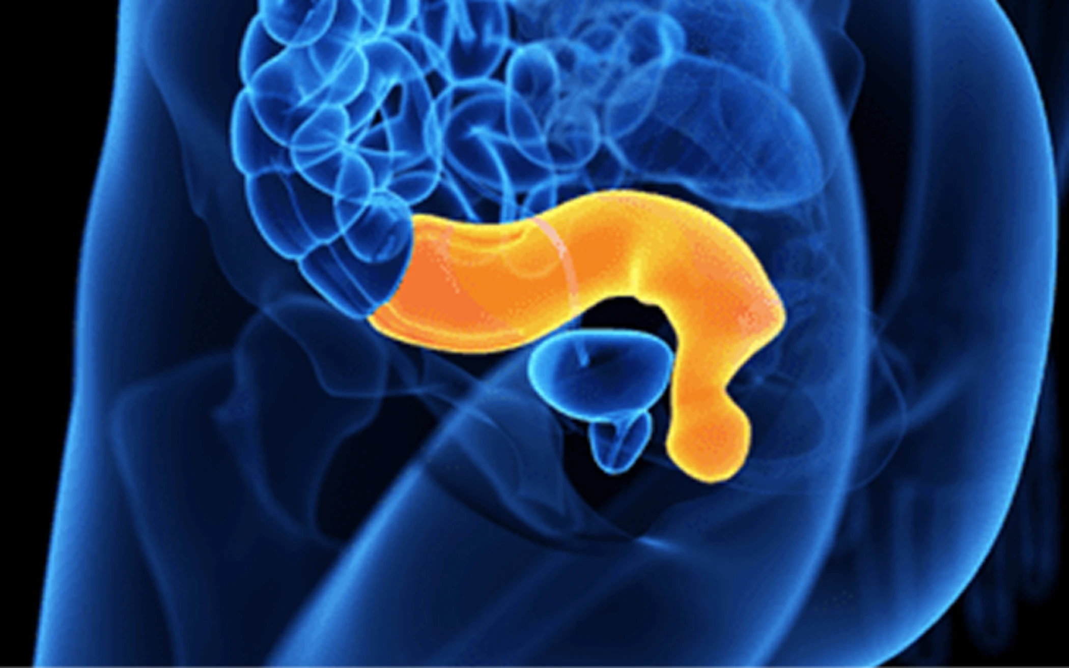
The rectum is the final segment of the large intestine that connects the colon to the anus. It stores waste produced in the colon until the body is ready to eliminate the waste through the process of defecation (having a poo!). It is a hollow muscular tube 6–8 inches (15–20 centimetres) in length and 2.5 inches in diameter at its widest point.
Although neuroendocrine cancer of the rectum is one of the most commonly diagnosed neuroendocrine cancers (in part related to colon cancer screening investigations) they are amongst the least common colorectal cancers (<2%).
In the early stage the neuroendocrine cancer of the rectum may present as a small polyp (less than 1–2 centimetres), or a flat doughnut shaped lesion. Larger polyps or lesions are possible, both with and without disease spread. In either case there are clear treatment and follow up protocols in place that will be discussed with you.
Symptoms
Symptoms of neuroendocrine cancer of the rectum may include:
- Bleeding from the bottom (rectal bleeding)
- Itching and pain around the rectum and or anus
- Small lumps around the anus
- A discharge of mucus
- Loss of bowel control (bowel incontinence)
- Constipation
- A feeling that your rectum doesn’t empty completely or a continual or recurrent feeling that you need to open your bowels (tenesmus)
- Weakness or fatigue
- Unexplained weight loss
However, many people experience no symptoms at all in the early stages of the disease. When symptoms appear, they may be related to the cancer’s size and location.
Neuroendocrine cancer of the rectum is rarely associated with a neuroendocrine associated syndrome (a syndrome is where two or more related symptoms occur).
Other, rarer symptoms, including paraneoplastic syndrome and oncological emergencies (a specific set of health concerns that can occur in any cancer), such as raised calcium levels (hypercalcaemia), may occur.
Tests
Tests that may be used for the diagnosis and/or monitoring of neuroendocrine cancer of the rectum:
Blood and urine
- Full blood count
- Vitamin B12 and serum iron
- Liver and kidney function
Biochemical:
- Chromogranin A (and B)
- Urinary 5-HIAA
- CEA
- β-HCG
- Pancreatic polypeptide
Scans and further investigations
- Endo-anal ultrasound
Sigmoidoscopy/colonoscopy with or without biopsy - Pelvic MRI
- Contrast CT of the chest/abdomen/pelvis
- Gallium-Dotatate PET/CT (SRS SPECT/CT if Dotatate PET not available)
- FDG-PET if high grade or rapidly progressing disease
Watch our video: Your Guide to Scans
Pathology (what can be seen through special tests under a microscope)
- Differentiation and cellular morphology
- Synaptophysin
- Chromogranin
- Ki67
- Low molecular weight keratins
- GLP
- PP/PYY
- CD56
- Prostatic acid phosphates
Grading
Not all cancers are the same or behave in the same way. Some may grow very slowly, may not have invaded nearby structures, or spread to other parts of the body at the time of diagnosis – but they have the potential to. However some may have already spread at the time of diagnosis. The grade is the rate at which the neuroendocrine cancer grows. You might see this referred to as ‘Ki67’ or ‘Mitotic Index’ mentioned in your clinic letters or medical reports. Grade 1 is slow growing through to grade 3, a more aggressive, rapidly growing disease.
Causes and risk factors
We do not know exactly what causes neuroendocrine cancer of the rectum, however research is ongoing to truly understand both the pathology and the biology with the hope that this will lead to a much better understanding of the causes.
Most neuroendocrine cancers do not run in families; however, there are a number of rare conditions that may increase your risk. Therefore, if other members of the family have been diagnosed with a particular cancer (especially aged 50 years or less) or cancer-associated genetic condition, it is important that you tell your specialist team about not only your medical history, but also any family medical illnesses or conditions.
How is neuroendocrine cancer of the rectum treated?
For all patients, there are many things to consider in planning treatments. Your treatment will be personalised to you and the type of neuroendocrine cancer you have. Even if you have a diagnosis that sounds the same as another patient, your treatment and follow up plan may be different.
Neuroendocrine cancer of the rectum is usually, but not exclusively, small and grade 1 or grade 2. Many may be completely removed, as a polyp, by an endoscopic resection. The size and stage of the neuroendocrine cancer is really important in deciding the correct treatment pathway.
Your care team will discuss your treatment options with you – giving you both written and verbal information – to help you make an informed choice. Together you can agree on the most appropriate treatment for you. There is consensus agreement that all neuroendocrine cancer patients should be reviewed by a specialist multidisciplinary team.
Treatment options will depend on the type, position and size of your neuroendocrine cancer, and whether (or to where) it has spread. It will also depend on whether you have any other health concerns and/or illnesses and your general health and fitness. A big part of meeting with your doctors or specialist nurse/team, is to make sure you get the information you need to understand your condition.
The key aim of treatment should be to help you have the best possible care and quality of life by ensuring access to appropriate treatment, management of symptoms and addressing what’s most important to you.
One or more of the approaches below may be suggested:
- Surgery
- Control of your disease, by slowing or stopping further growth and/or spread
- Palliation, or easing, of any symptoms you may be experiencing
Surveillance
Monitoring through clinic review, bloods and scans can be used to assess how well treatment is working or in periods between treatments (which may be months or even years).
As not everyone will need to be on treatment, surveillance can be used to check your cancer and general health for any signs of change that may mean that a treatment might need to be considered. All treatments have possible side effects, therefore, it is important to know when treatment may be helpful for you or not.
Surgery
Surgery will depend on site, size, position and spread of your neuroendocrine cancer. Some rectal cancers develop within polyps; others form solid tumours.
A polyp is a small cell clump that develops in two ways. Pedunculated polyps hang from a short stalk. Sessile polyps are flat and grow directly out of the surrounding tissue.
If your neuroendocrine cancer develops within a polyp you may be able to have it removed by:
Polypectomy
Polyps are removed during endoscopy using hot or cold forceps or a snare.
- Endoscopic mucosal resection (EMR): an endoscope is introduced to the site of the polyp. Using a small needle a fluid is injected underneath to raise the polyp creating a safe field to remove it using a snare. The area is then cauterised (to stop bleeding).
- Endocopic submucosal dissection (ESD): the procedure is similar to EMR but uses a small knife, rather than a snare, to remove the polyp, cauterising blood vessels at the same time to prevent bleeding.
If it is a large polyp or small tumour you may be able to have it removed by:
- Transanal mucosal surgery (TEMS): using a specially designed microscope and instruments, this procedure allows surgery to be performed through the anus (back passage) inside the rectum.
- Transanal minimally invasive surgery (TAMIS): a specialised laparoscopic surgery port is placed into the anus. The laparoscope and instruments required to carry out the procedure are passed through the port to the rectum, so that the polyp or tumour can be surgically removed.
Larger or more invasive disease will require more complex surgery:
- Total mesorectal excision (TME): surgery to remove the rectum and sigmoid colon, including the blood supply and associated lymph glands. This procedure may result in a temporary or permanent colostomy dependent on whether is possible to rejoin the end of the healthy bowel to the anus.
- Anterior-perineal extirpation: surgery to remove the lower part of your (descending) colon, sigmoid, rectum and anus – along with associated lymph glands. This procedure results in a permanent colostomy.
A colostomy is formed by bringing part of your colon (large bowel) or the end or loop of ileum (small bowel) out on to the surface of your abdomen to form a stoma (an opening) – and may be temporary or permanent – depending on how much bowel is removed, how much you have left and how healthy your remaining bowel is. Waste passes out of the stoma and is collected in an external pouch (known as a stoma bag). If your colostomy is temporary, further surgery may take place at a later date, to rejoin your bowel and close the stoma – this is called a stoma reversal.
Non-surgical treatments
- Somatostatin analogues (SSAs)
- Chemotherapy
- Targeted molecular therapies
- Peptide receptor radionuclide therapy (PRRT)
- Radiotherapy
- Interventional radiology
- Clinical trials
- Symptom control
Diagnosis and living with neuroendocrine cancer of the rectum
A diagnosis of neuroendocrine cancer can bring questions, uncertainty and a range of emotions. Information and support are available to help you understand your diagnosis and navigate what happens next.
Follow up
There are expert agreed guidelines regarding how and when follow up should occur, however, in practice this varies and often with good reason. Follow up should be expert informed and evidence/research based but also tailored to you and what is appropriate for your best care.
Following surgery/endoscopic resection – for minimum 10 years:
- G1/2 <1 cm: no lymph nodes, no evidence of invasion: there is no data re recommended follow up
- G3 <1 cm: annual follow up with colonoscopy
- G1/2 1-2 cm: annual follow up with colonoscopy
- Biochemistry alongside colonoscopy/scan
All neuroendocrine cancers >2 cm require ongoing follow up – minimum 10 years (NB metastatic disease has been seen beyond this time):
- G1/2: annual colonoscopy + CT + biochemistry
- G3: 4 to 6 monthly colonoscopy + CT + biochemistry for the first year, then annually
If liver tumours are present, MRI or contrast CT is performed concurrent with follow up timings.
How you can be part your care
You are a key part of your team. You can help by:
- Reporting new symptoms or any changes in current symptoms or overall health
- Asking questions if at all uncertain or you want more information about your diagnosis and/or plan of care
- Attending appointments made – and checking up with the team if you haven’t received an appointment according to your agreed plan of care.
- Let your team know of your preferences and wishes regarding care – what is most important to you.
Advanced disease
Follow up should be aligned with guidelines, and also guided by prognosis, expected treatment efficacy and treatment related toxicity. Your health, wellbeing, physical activity, informed choice and preference for ongoing care as well as the aim of treatment should be reviewed and discussed to best plan care.
Stories from our community
Hear from people connected to neuroendocrine cancer as they share their experiences, perspectives and insights – from living with the condition to caring for, treating and supporting those affected.
Remember
Remember: every person’s experience with neuroendocrine cancers of the rectum is different – there may be shared similarities, but each journey is unique.
Your healthcare team will support you every step of the way, guiding your treatment, care, and follow-up. We’re here alongside them, offering additional support and information whenever you need it.
The information on this page is based on available clinical expert guidelines and publications. It is for informational purposes only and should not be considered professional medical advice.