
Thyroid

Find information and support for people affected by medullary thyroid cancer (MTC), including links to further resources.
Thymus

Neuroendocrine cancer of the thymus is very rare and often causes no symptoms at first. Others develop a cough, chest pain or shortness of breath, and occasionally the cancer produces excess hormones, adding further symptoms to look out for.
Male reproductive system

Neuroendocrine cancer of the testes is very rare. It may cause a lump, swelling or discomfort in a testicle, but is often found during investigations or surgery for suspected testicular cancer rather than from symptoms alone.
Stomach

Neuroendocrine cancer of the stomach is rare and can affect people in different ways, depending on the type of cancer.
Small bowel

Neuroendocrine cancer of the small bowel often causes no symptoms at first. Others have abdominal pain, changed bowel habits or symptoms of carcinoid syndrome, and sometimes it is only found after a bowel obstruction or tests for something else.
Skin

Merkel cell carcinoma is a rare neuroendocrine cancer of the skin: a firm, painless lump that can grow quickly, usually on sun-exposed skin. It accounts for about a third of rare skin cancers, with over 35% arising on the head and neck.
Rectum
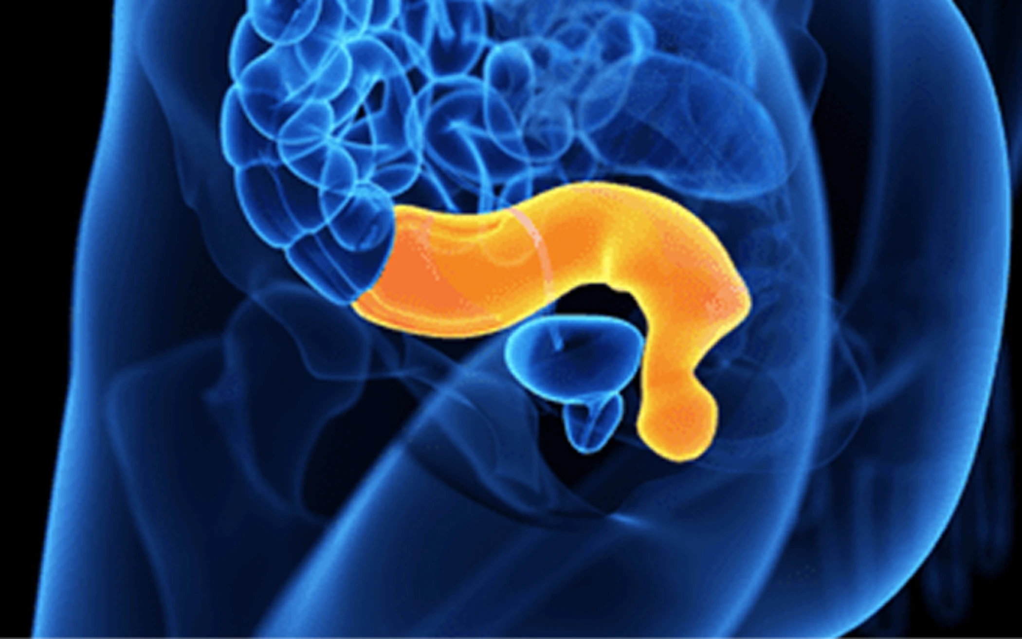
Neuroendocrine cancer of the rectum is often found during a routine bowel investigation and may cause no symptoms at all. The type, size and grade of the cancer, and whether it has spread, guide the treatment you are offered.
Paraganglioma

Paraganglioma is a rare neuroendocrine tumour that develops from specialised cells near nerves and blood vessels, most often in the abdomen. Functioning tumours release hormones such as adrenaline, causing headaches, sweating and palpitations.
Pancreas

Pancreatic neuroendocrine cancer can produce excess hormones in some people and tumour-related symptoms in others. It can also cause pancreatic exocrine insufficiency, with pale fatty stools, weight loss and bloating, managed with enzyme replacement therapy.
Lung

Large cell neuroendocrine carcinoma (LCNEC) of the lung is a rare, faster-growing type of neuroendocrine cancer that develops in the lungs. It is one of two main types of lung neuroendocrine carcinoma (NEC), alongside small cell lung cancer (SCLC).